-
The University
- Welcome
- Who we are
- Media & PR
-
Studying
- General
- Degree programs
- Campus life
-
Research
- Profile
- Infrastructure
- Cooperations
- Services
-
Career
- Med Uni Graz as an Employer
- Educational Opportunities
- Work Environment
- Job openings
-
Diagnostics
- Patients
- Referring physicians
- Health Topics
- Health Infrastructure

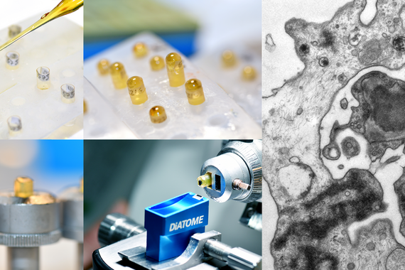
Electron microscopy
In the electron microscopy lab, a tissue sample is irradiated with electrons under high voltage in a vacuum in order to depict the inside or surface of cells. The electron microscope reaches a significantly higher resolution (currently about 0.1 nm) than an optical microscope (about 200 nm).

This method is applied to assess ultrastructures such as organelles or immune complexes that cannot be detected with an optical microscope.
The tissue sample is processed in special reagents and must first be cut into semi-thin sections (about 0.5–1µm) and then into ultra-thin sections (<100nm). This highly specialized procedure takes several days to complete.
Electron microscopy tests are an important tool in diagnosing kidney disease, which is the most frequent indication for the use of electron microscopy at our institute (up to 80%). Other diseases such as muscular disorders, ciliopathies, metabolic disorders, neurodegenerative diseases and hereditary skin and connective tissue diseases are investigated. The electron microscope is also used to solve scientific problems.
Diese Methode wird insbesondere zur Beurteilung lichtmikroskopisch nicht erkennbarer Ultrastrukturen wie z.B. Zellorganellen oder Immunkomplexen angewandt.
Die Aufarbeitung der Gewebeprobe erfolgt in speziellen Reagenzien und erfordert zuerst ein Schneiden in Semidünnschnitte (etwa 0,5-1µm) und danach in Ultradünnschnitte (<100nm). Dieser hochspezialisierte Verarbeitungsprozess dauert mehrere Tage.
Elektronenmikroskopische Untersuchungen sind ein wesentliches Instrument in der Diagnosestellung von Nierenerkrankungen, welche mit bis zu 80% die häufigste Indikation zur Elektronenmikroskopie an unserem Institut darstellen. Zusätzlich werden andere Erkrankungen, wie Muskelerkrankungen, Ziliopathien, Stoffwechselerkrankungen, neuro-degenerative Erkrankungen und erblich bedingte Haut- und Bindegewebserkrankungen untersucht. Zudem wird das Elektronenmikroskop für wissenschaftliche Fragestellungen eingesetzt.
Lab
Medical division management
Deputy division management